Toll Free: (888) 496-4400
Main Office: (801) 264-4444
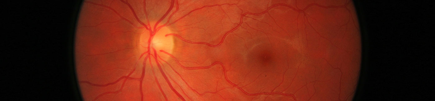
Retinal Detachment
There are 3 kinds of retinal detachments: (1) Rhegmatogenous, (2) Tractional, and (3) Exudative.
A rhegmatogenous retinal detachment results when a tear or break in the retina occurs, resulting in vitreous fluid gaining access to the subretinal space. The accumulation of fluid in the subretinal space results in the separation of the retina from the retinal pigment epithelium.
Most detachments generally are of the rhegmatogenous type. They occur with an incidence of 5-10 per 100,000 population/year. They tend to be more common in men. Any retinal break or tear can cause a retinal detachment. Studies have reported that approximately 6% of all eyes have a retinal break, but only 1 person in 10,000-15,000 a year eventually requires retinal detachment repair.
A tractional retinal detachment is caused by direct mechanical traction. These detachments occur when scar tissue contracts and induces mechanical traction on the neurosensory retina, thus pulling the retina from the retinal pigment epithelium.
An exudative retinal detachment occurs in the absence of either retinal breaks or vitreoretinal traction. These detachments result from diseases of the retinal pigment epithelium and the choroids. The retinal pigment epithelium is responsible for pumping fluid out of the retina so that the retina and retinal pigment epithelium remain attached. Any disease of the retinal pigment epithelium and the choroid can damage the blood retinal barrier and overwhelm the retinal pigment epithelium pump, resulting in fluid accumulating under the retina. Examples include inflammation, vascular injury, and choroidal tumors.
Treatment
Pneumatic retinopexy, scleral buckling, and vitrectomy are established modalities of treatment for retinal detachments. Each procedure is different and is selected on a case-by-case basis, depending on the patient and the location of the retinal break(s) that result in the retinal detachment.
Pneumatic retinopexy is an in-office procedure that involves injection of an inert gas into the vitreous cavity. This procedure is selected for retinal breaks that are found in the superior retina. The retinal break is usually treated with cryotherapy, followed by injection of the inert gas to tamponade and close the retinal break. The gas bubble is slowly reabsorbed by the body and does not need to be removed by additional procedures.
Scleral buckling surgery has been the most common surgical method employed in repairing retinal detachments. This procedure has been utilized for nearly five decades. Scleral buckling involves the placement of a silicone band that is sutured to the external sclera in order to indent the eye and reduce the retina to retinal pigment epithelium distance. The indentation results in closure of the retinal break, allowing for reabsorption of the subretinal fluid by the retinal pigment epithelium.
Pars plana vitrectomy is a surgical procedure that uses microinstruments to remove the vitreous gel that is responsible for the traction on the retinal tears. A vitrectomy is advantageous in retinal detachments that have vitreous hemorrhage, precluding a view of the retina. After the vitreous gel is removed, the retinal tear is treated with cryotherapy or laser eye surgery, followed by instillation of an inert gas or silicone oil in the vitreous cavity to tamponade the retinal break(s).
The decision to perform one surgery over another should be discussed extensively by the retina specialist as each procedure has advantages and disadvantages that are specific for every patient.